Electron-Based Imaging Technologies
The combination of magnetism with nanostructures opens the possibility to exploit the electrons’ spin degree of freedom for so-called spintronic devices with optimized properties or new functionalities. Over the last years, the group has focused on the development of new instruments based on the use of electrons for imaging of spintronic devices, which operate at the highest spatial resolution.
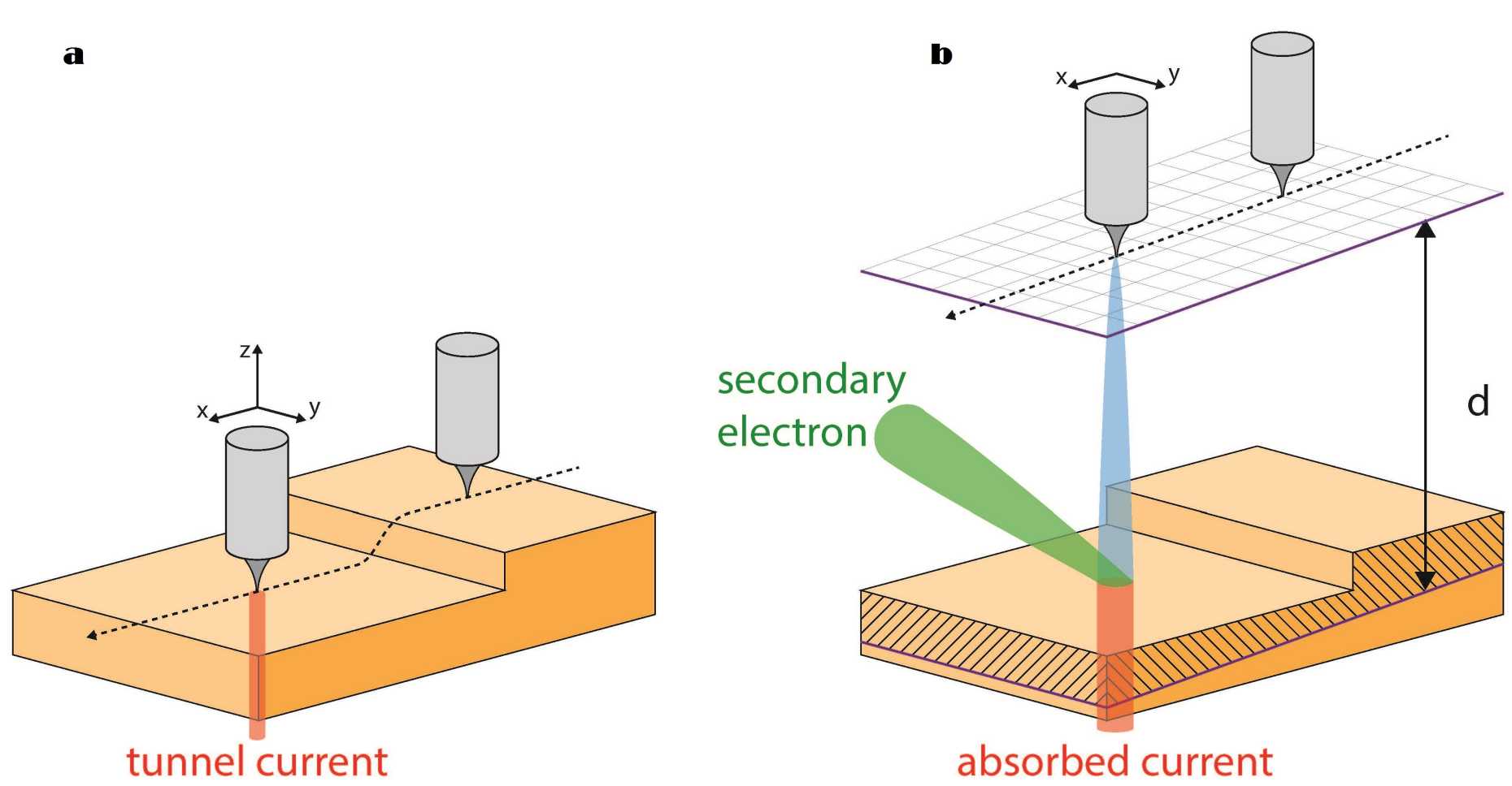
One example of such an instrument is the Scanning Field Emission Microscope (SFEM), which combines the high spatial resolution of Scanning Tunneling Microscopy (STM) with the spectroscopic and spin analysis capabilities of secondary electrons emitted in conventional Scanning Electron Microscopy. In a first step (see (a) in the Figure), the tip (grey) is approached to the surface (in the present case, two “yellow” terraces separated by a monoatomic step) at subnanometre distances (STM mode). In STM, transport of electrons between tip and surface occurs by direct (Binnig–Rohrer) quantum mechanical tunneling and allows the recording of a topographic map of the surface with atomic spatial lateral resolution. After the acquisition of such an STM image, the tip is retracted along the z-axis away from the surface up to distances d of about 10 nanometres (b, SFEM mode). At these distances, direct quantum mechanical tunneling is entirely suppressed, but electrons can be field emitted from the tip. The field-emitted electrons (blue in (b)) are accelerated towards the surface of the target. Upon striking the target, they are partly absorbed (red in (b)) but also generate (by virtue of elastic and inelastic scattering processes) a cascade of secondary backscattered electrons (green in (b)). Those secondary backscattered electrons that escape the tip-target junction and reach the macroscopic environment surrounding the junction can be analyzed according to their intensity, producing a “secondary electron image'' of the topography of the target itself. Alternatively, they can be selected according to their energy, enabling spectro-microscopy. Or, else, their spin polarization can be analyzed, producing an image of the magnetic contrast of the target. In any case, the “simple” operation of retracting the tip “a little bit “ away from the target turns the STM instrument into a lensless low energy Scanning Electron Microscope with the possibility of secondary electron imaging and spin analysis.